Knee Muscle Anatomy Mri ~ Case Study Medial Meniscal Repair And Chondroplasty Of The Right Knee In A 50 Year Old Male. This allows them to inspect the elements of the. The knee joint is a complex joint that connects three bones; Mri patterns of neuromuscular disease involvement thigh & other muscles 2. There is a wide variety of variant vascular anatomy and vascular pathology that can occur around the knee, including an aberrant anterior tibial artery, vascular trauma that occurs with knee dislocation, popliteal artery entrapment syndrome, popliteal artery aneurysm, popliteal vein thrombosis, cystic adventitial. General anatomy and musculoskeletal system.
The muscles of the knee include the quadriceps, hamstrings, and the muscles of the calf. Please email baodo at stanford.edu This mri knee sagittal cross sectional anatomy tool is absolutely free to use. 12 photos of the knee muscle anatomy mri. Scroll through the structures to understand the anatomy.
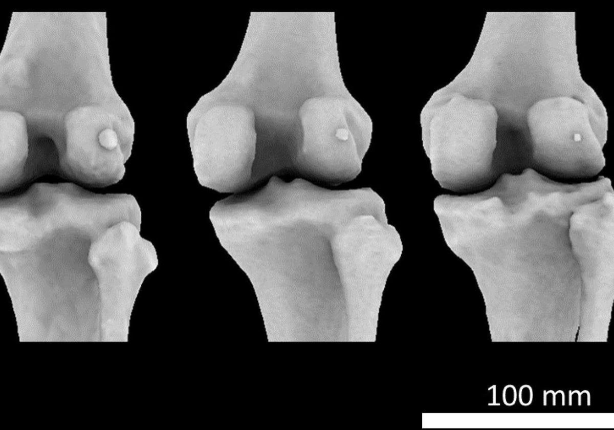
Tiny Knee Bone Once Lost In Humans Is Making A Comeback The Scientist Magazine from cdn.the-scientist.com Radiologists must familiarize themselves with typical mri findings to accurately detect and classify muscle injuries. Stanford bone tumor ddx | iss/ssr msk lectures | ocad msk cases stanford msk mri atlas has served over 1,000,000 pages to users in over 100 countries. Thigh muscles also protect neurovascular structures as they go through the proximal hip joint to the knee and lower leg (3). Learn about the muscles, tendons, bones, and ligaments that comprise the knee joint anatomy. Rubin da, kettering jm, towers jd, britton ca: The knee is designed to fulfill a number of functions: The femur, tibia and patella.the arrangement of the bones in the knee joint, along with its many ligaments, provide it with the arthrokinematics that allows for great stability, combined with great mobility.being arguably the most stressed and exposed joint of the body, the knee joint is predisposed to various. These motions of the knee allow the body to perform such important movements as walking, running, kicking, and jumping.
The knee is designed to fulfill a number of functions:
Stanford bone tumor ddx | iss/ssr msk lectures | ocad msk cases stanford msk mri atlas has served over 1,000,000 pages to users in over 100 countries. Popliteus muscle popliteus tendon posterior horn of lateral meniscus head of fibula anterior horn of lateral meniscus lateral femoral condyle 58. The muscles that affect the knee's movement run along the thigh and calf. Knee anatomy francesc malagelada jordi vega pau golanó the knee is the largest joint in the human body and one of the most complex from a functional point of view. Magnetic resonance imaging is particularly well suited for the medical evaluation of the musculoskeletal (msk) system including the knee, shoulder, ankle, wrist and elbow. Articular surface of patella and femur, condyle, epicondyle and muscles (popliteus, sartorius, gastrocnemius, semimembranous with tendos.) the images obtained were exported to jpeg from dicom data stored on the pacs (picture archiving and communicating system). Medical images from an mri allow medical professionals to distinguish body tissues, including the meniscus (shock absorbers in the knee), cartilage, tendons, and ligaments. Medically reviewed by stuart hershman, md What an mri can tell you about a knee injury. The knee joins the thigh bone (femur) to the shin bone (tibia). 12 photos of the knee muscle anatomy mri. Scroll through the structures to understand the anatomy. In conclusion, we describe the normal mri anatomy of the distal biceps femoris and the relationship of this muscle with the common peroneal nerve.
The smaller bone that runs alongside the tibia (fibula) and the kneecap (patella) are the other bones that make the knee joint. The common peroneal nerve typically courses downward within abundant fat posterior to the short head of the biceps femoris muscle and superficial to the lateral head of the gastrocnemius muscle, but. Rubin da, kettering jm, towers jd, britton ca: Louis, usa and the rijnland hospital in leiderdorp, the netherlands The test can show a range of.
Jaypeedigital Ebook Reader from d45jl3w9libvn.cloudfront.net Radiology imaging medical imaging subscapularis muscle shoulder anatomy bicep tendonitis mri brain shoulder rehab rotator cuff tear anatomy this mri knee cross sectional anatomy tool is absolutely free to use. The muscles of the knee include the quadriceps, hamstrings, and the muscles of the calf. Injuries such as anterior cruciate ligament, meniscus and rotator cuff tears are all easily diagnosed when there is a firm understanding and knowledge of human anatomy. Magnetic resonance imaging (mri) is the test of choice to confirm the diagnosis of a torn meniscus. The muscles that affect the knee's movement run along the thigh and calf. Magnetic resonance imaging (mri) is the highest standard in diagnostic imaging of muscle injuries(1). Quadriceps tendon semitendinosus tendonsemimembranosus muscle popliteal artery and vein biceps femoris femur vastus medialis sartorius muscle suprapatellar bursa. Mri of the knee is often performed for presumed musculoskeletal conditions.
Magnetic resonance imaging is particularly well suited for the medical evaluation of the musculoskeletal (msk) system including the knee, shoulder, ankle, wrist and elbow.
Learn about the muscles, tendons, bones, and ligaments that comprise the knee joint anatomy. Medical images from an mri allow medical professionals to distinguish body tissues, including the meniscus (shock absorbers in the knee), cartilage, tendons, and ligaments. Doctors may recommend a knee mri if a patient experiences the following(3): Rubin da, kettering jm, towers jd, britton ca: An exercise program can strengthen the muscles surrounding the knee, increasing the knee's stability. A knee mri looks specifically at your knee and its surrounding areas. The knee joint is the junction of the thigh and leg. Please email baodo at stanford.edu Atlas of knee mri anatomy this webpage presents the anatomical structures found on knee mri. Knee anatomy francesc malagelada jordi vega pau golanó the knee is the largest joint in the human body and one of the most complex from a functional point of view. This mri knee sagittal cross sectional anatomy tool is absolutely free to use. An mri lets your doctor see the soft tissues in your body along with the bones. Radiologists must familiarize themselves with typical mri findings to accurately detect and classify muscle injuries.
12 photos of the knee muscle anatomy mri. What an mri can tell you about a knee injury. Rubin da, kettering jm, towers jd, britton ca: The muscles that affect the knee's movement run along the thigh and calf. These muscles work in groups to flex, extend and stabilize the knee joint.
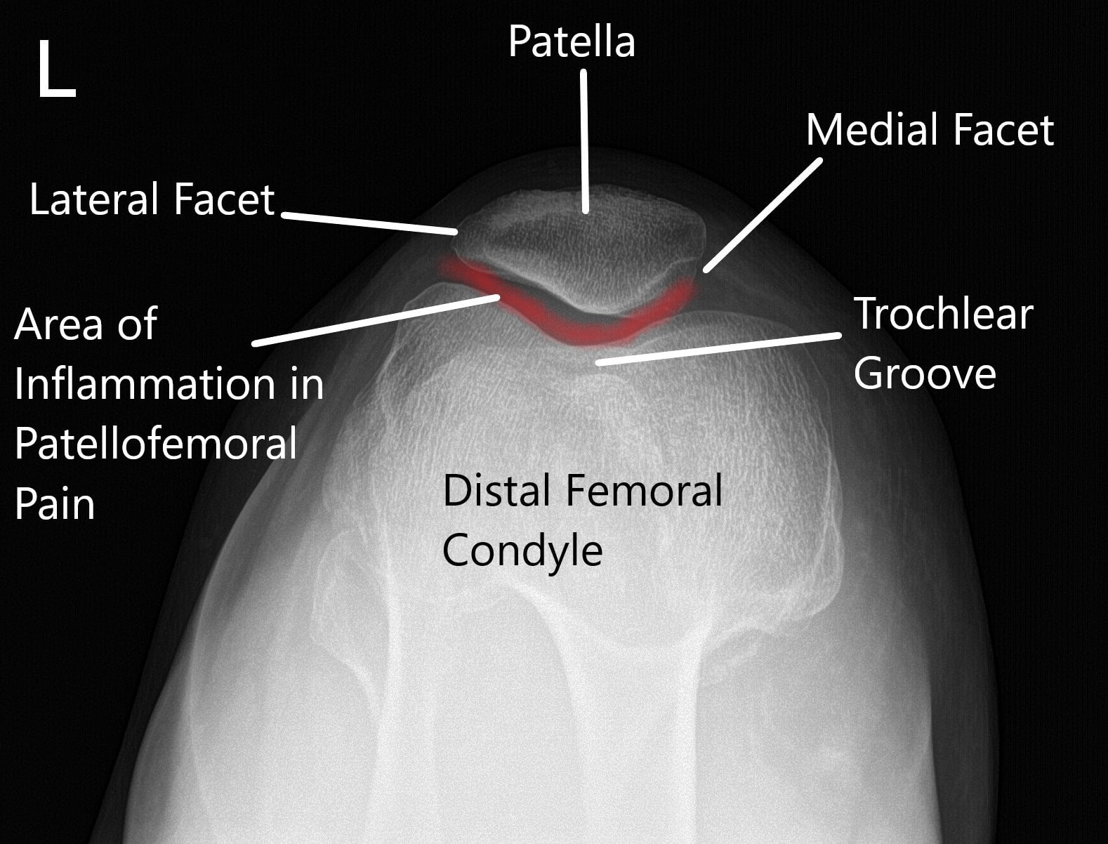
Runners Knee New York Dr Nakul Karkare from www.cortho.org Knee mri scan from www.ucsfhealth.org an understanding of normal anatomy and biomechanics of the knee extensor mechanism is necessary to comprehend the imaging of extensor mechanism injuries. Injuries of the patellofemoral joint. Magnetic resonance imaging (mri) is the test of choice to confirm the diagnosis of a torn meniscus. 12 photos of the knee muscle anatomy mri. Rubin da, kettering jm, towers jd, britton ca: Atlas of knee mri anatomy this webpage presents the anatomical structures found on knee mri. The knee is designed to fulfill a number of functions: Knee anatomy francesc malagelada jordi vega pau golanó the knee is the largest joint in the human body and one of the most complex from a functional point of view.
Mri knee joint anatomy 1.
Learn about the muscles, tendons, bones, and ligaments that comprise the knee joint anatomy. The test can show a range of. Please email baodo at stanford.edu The muscles of the knee include the quadriceps, hamstrings, and the muscles of the calf. A knee mri looks specifically at your knee and its surrounding areas. Magnetic resonance imaging is particularly well suited for the medical evaluation of the musculoskeletal (msk) system including the knee, shoulder, ankle, wrist and elbow. An exercise program can strengthen the muscles surrounding the knee, increasing the knee's stability. Diagnosing muscle injuries of the hip and thigh is a relevant issue in professional sports(2). Home » unlabelled » knee muscle anatomy mri : These muscles work in groups to flex, extend and stabilize the knee joint. Stanford bone tumor ddx | iss/ssr msk lectures | ocad msk cases stanford msk mri atlas has served over 1,000,000 pages to users in over 100 countries. Mri of the knee is often performed for presumed musculoskeletal conditions. Injuries of the patellofemoral joint.
Share :
Post a Comment
for "Knee Muscle Anatomy Mri ~ Case Study Medial Meniscal Repair And Chondroplasty Of The Right Knee In A 50 Year Old Male"


Post a Comment for "Knee Muscle Anatomy Mri ~ Case Study Medial Meniscal Repair And Chondroplasty Of The Right Knee In A 50 Year Old Male"